Breast Cancer – Causes, Symptoms, Treatments
|
Reading time: 12 minutes
|
Key Takeaways
- Breasts are composed of fatty tissue, lobes, ducts, and lymph nodes; mutations lead to cancerous growths.
- Types of Breast Cancer include in situ (confined to ducts/lobules) and invasive (spreads beyond ducts/lobules) which includes ductal and lobular carcinoma.
- Symptoms include lump, changes in size/shape, nipple discharge, skin changes, and breast pain/tenderness.
- Risk Factors include non-modifiable (age, genetics) and modifiable (hormonal factors, lifestyle choices) contribute to susceptibility.
- Diagnosis and treatment include physical exams, imaging tests (mammograms, ultrasounds, MRIs), biopsies, surgery (lumpectomy, mastectomy), systemic treatments (chemotherapy, hormone therapy), and targeted therapy are utilized.
What is Breast Cancer?
Breasts are made up of fatty tissue, connective tissue, lobes (milk-producing glands), ducts (tubes that carry milk to the nipple), and lymph nodes (part of the immune system). Breast cancer starts when normal cells in the breast mutate and become cancerous. These cells multiply rapidly and form tumors. There are two main categories.
In situ carcinoma is an early stage where cancer cells are confined to the milk ducts (ductal carcinoma in situ – DCIS) or lobules (lobular carcinoma in situ – LCIS) and haven’t invaded surrounding tissues. It’s not life-threatening but can progress to invasive cancer.
With invasive carcinoma, cancer cells break through the ducts or lobules and invade surrounding breast tissue. This has a higher risk of spreading (metastasizing) to lymph nodes and other parts of the body.
Types of Invasive Breast Cancer:
Invasive ductal carcinoma (IDC), also sometimes called infiltrating ductal carcinoma, is the most common form of breast cancer, accounting for roughly 75-80% of all breast cancer diagnoses. It starts in the milk ducts of the breast, the tubes that carry milk from the lobules (milk-producing glands) to the nipple.
Abnormal cells grow and invade the surrounding breast tissue beyond the walls of the duct. At this stage, the cancer cells have the potential to spread further through the lymph nodes or bloodstream to other parts of the body. This is called metastatic breast cancer.
Invasive lobular carcinoma (ILC), also known as infiltrating lobular carcinoma, is the second most common type of invasive breast cancer, accounting for around 10-15% of all diagnosed cases. Unlike IDC, which originates in the milk ducts, ILC starts in the lobules, the milk-producing glands of the breast.
Invasive lobular carcinoma cells tend to spread in a single-file line or in small clusters throughout the breast tissue, making them sometimes harder to detect on mammograms compared to IDC which forms a lump. It is generally considered slower-growing than IDC, but it can still be aggressive and spread to other parts of the body.
Over 80% of ILC cases are estrogen receptor-positive (ER+) and HER2-negative (HER2-), meaning they are fueled by the hormone estrogen and don’t overexpress the HER2 protein. This can influence treatment options.
Less Common Types of Invasive Breast Cancers
Less common types of invasive breast cancers include Inflammatory breast cancer, Paget’s disease of the breast, and metaplastic breast cancer.
Inflammatory breast cancer (IBC) is a rare and aggressive form of breast cancer, accounting for only 1-5% of all breast cancers. It differs from other breast cancers in its presentation and behavior.
Unlike typical breast cancers that cause lumps, IBC disrupts the lymphatic system in the breast. These cells block the lymph vessels in the breast, preventing fluid drainage. This buildup of fluid leads to swelling and redness, mimicking inflammation, with additional symptoms like the affected area being overly warm when touched, and a texture resembling an orange peel on the affected breast.
IBC is fast-growing and can spread quickly to the lymph nodes and other parts of the body. Early detection is crucial for successful treatment. However, since it/ presents like an infection, it can be misdiagnosed, delaying proper treatment.
Symptoms of Breast Cancer
Breast cancer symptoms can vary from person to person, and some women might not experience any symptoms at all. However, here are some common warning signs to be aware of.
- A lump or mass in the breast or underarm area: This is often the most noticeable symptom. However, not all lumps are cancerous, but it’s essential to have any new lump or mass evaluated by a healthcare professional.
- Changes in breast size or shape: Any noticeable changes in the size or shape of the breast, including swelling, asymmetry, or distortion, should be examined.
- Changes in the appearance of the breast or nipple: This may include dimpling or puckering of the breast skin, redness or scaling of the nipple or breast skin, or nipple inversion (turning inward).
- Nipple discharge: Spontaneous nipple discharge, particularly if it’s bloody or clear, may be a sign of breast cancer. However, many benign conditions can also cause nipple discharge.
- Breast pain or tenderness: While breast pain is not typically a common symptom of breast cancer, it can occur in some cases. However, it’s essential to note that most breast pain is not associated with cancer.
- Changes in the texture or color of the breast skin: This may include thickening or dimpling of the skin, resembling the texture of an orange peel, or changes in the skin color or texture.
Causes of Breast Cancer
The exact cause of breast cancer isn’t fully understood, but research has identified several factors that increase the risk of developing it. Inherited mutations in certain genes, like BRCA1 and BRCA2, significantly elevate breast cancer risk. These genes play a role in DNA repair, and when mutated, they can leave cells vulnerable to damage that can lead to cancer.
Exposure to estrogen over a longer period can increase risk. This includes factors like starting menstruation at a young age, having late menopause (after age 55), and taking hormone replacement therapy (HRT) for an extended period.
Lifestyle Factors such as obesity, especially after menopause, can also increase the risk of developing breast cancer. Fat tissue produces estrogen, which can fuel the growth of some breast cancers. Lack of physical activity and excessive alcohol consumption is another potential risk factor.
Risk Factors That Increase the Chance of Developing Breast Cancer
There are several factors that can increase a woman’s risk of developing breast cancer. These can be broadly categorized into modifiable and non-modifiable factors.
Non-Modifiable Risk Factors:
- Age: Risk increases as women age, with most cases diagnosed after age 50.
- Family History: Having close relatives (mother, sister, daughter) with breast cancer significantly increases your risk.
- Genetics: Inherited gene mutations, particularly BRCA1 and BRCA2, significantly elevate breast cancer risk.
- Dense Breast Tissue: Having denser breast tissue can make mammograms less effective in detecting cancer and might be a risk factor itself.
Modifiable Risk Factors:
- Hormonal Factors:
- Menstrual History: Starting menstruation at a young age (before 12) and going through menopause later in life (after 55) exposes a woman to estrogen for a longer period, increasing risk.
- Hormone Replacement Therapy (HRT): Some types of HRT, particularly those combining estrogen and progesterone used for more than five years, can slightly elevate risk.
- Lifestyle Factors:
- Weight: Being overweight or obese, especially after menopause, increases risk as fat tissue produces estrogen.
- Physical Inactivity: Lack of exercise is associated with a higher risk of breast cancer.
- Alcohol Consumption: Excessive alcohol intake can be a risk factor.
Other Potential Risk Factors:
- Radiation Exposure: Exposure to ionizing radiation, particularly at a young age, can increase risk. However, the amount of radiation from mammograms is very low and not considered a significant concern.
- Night Shift Work: Studies suggest disruptions in circadian rhythms due to night shift work might influence hormone levels and increase risk, but more research is needed.
How is Breast Cancer Diagnosed?
Diagnosing breast cancer typically involves a multi-step process. During a physical exam, a doctor will palpate your breasts, feeling for lumps, changes in size or shape, dimpling, or nipple retraction/inversion. They will also examine your lymph nodes, checking for enlargement or tenderness, particularly in the armpits and near the collarbone, as these are the first places breast cancer might spread. This allows the doctor to identify any abnormalities that warrant further investigation.
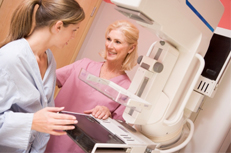
Once a physical exam raises concerns, imaging tests provide further confirmation of suspected cases. Mammograms are the primary screening tool for breast cancer, especially for women over 40. It uses low-dose X-rays to create images of breast tissue, revealing potential issues like lumps or masses, microcalcifications (tiny clusters of calcium deposits), and changes in breast tissue density. Both conventional and digital mammograms use X-rays to capture an image of your breast tissue. However, the key difference lies in how the image is stored:
- Conventional Mammography: Here, the X-ray image is recorded directly on film. This film needs physical storage and can be less convenient for sharing with other healthcare providers.
- Digital Mammography: This method creates an electronic image of your breast tissue. The image is stored as a computer file, allowing for easier storage, retrieval, and sharing with specialists.
Digital mammography offers several advantages over the conventional method:
- Electronic Storage and Sharing: Images are readily available on computers, facilitating evaluation and sharing between healthcare providers involved in your care.
- Multiple Images for Better Views: Typically, two X-ray images are captured from different angles (top-to-bottom and side-to-side) to provide a more comprehensive two-dimensional (2D) view of the breast tissue.
- 3D Mammography (Tomosynthesis): This advanced technique involves taking multiple low-dose X-ray images of the breast from different angles as the machine scans in an arc. These images are then reconstructed by a computer to create a three-dimensional (3D) view of the breast tissue. This 3D view can potentially provide a clearer picture and improve cancer detection, especially in women with dense breast tissue.
Ultrasounds are another key component of the diagnosis process. This test uses sound waves to create real-time images of the breast, helpful in differentiating between solid lumps and fluid-filled cysts. It can also be used to guide needle biopsies.
Magnetic Resonance Imaging (MRI) is another standard test used. It creates detailed internal pictures of the breast using strong magnets and radio waves. It is often used for women with high breast cancer risk due to family history or genetic mutations, women with dense breast tissue, or inconclusive mammogram results.
If a lump or abnormality is detected during an exam or imaging test, a biopsy is usually needed to confirm the presence or absence of cancer cells.
- There are different types of biopsies:
- Fine-needle aspiration (FNA): A thin needle is inserted to withdraw a small amount of cells for analysis. It’s less invasive but may not always provide enough tissue for conclusive diagnosis.
- Core needle biopsy: A thicker needle removes a core of tissue for more detailed examination.
- Surgical biopsy: In some cases, a small lump or suspicious area may be surgically removed for analysis.
Once cancer is diagnosed, further tests may be done to determine the stage and characteristics of the cancer, such as hormone receptor status (estrogen and progesterone receptor tests) and HER2 protein testing. This information helps guide treatment decisions.
How is Breast Cancer Treated?
Breast cancer treatment is multifaceted and depends on several factors, including the stage and type of cancer, your overall health, and your preferences.
- Local Treatments:
- These treatments aim to remove or destroy cancer cells in the breast and surrounding tissues:
- Surgery: The most common form of local treatment. Different types of surgery may be performed depending on the extent of cancer:
- Lumpectomy: Removal of the cancerous tumor with a small margin of healthy tissue around it. This is often followed by radiation therapy.
- Mastectomy: Removal of the entire breast. There are different types of mastectomies, including removing only the skin and nipple (skin-sparing mastectomy) or removing the entire breast and chest wall muscles (modified radical mastectomy).
- Sentinel lymph node biopsy: Removal of the first lymph nodes to which cancer is most likely to spread. If these nodes are free of cancer, no further lymph node removal may be needed.
- Axillary lymph node dissection: Removal of some or all lymph nodes in the armpit to check for cancer spread.
- Surgery: The most common form of local treatment. Different types of surgery may be performed depending on the extent of cancer:
- These treatments aim to remove or destroy cancer cells in the breast and surrounding tissues:
- Systemic Treatments:
- These medications travel through the bloodstream to target cancer cells throughout the body, potentially eliminating microscopic disease or preventing recurrence:
- Chemotherapy: Uses powerful drugs to kill cancer cells that may have spread beyond the breast. Chemo can be given before surgery (neoadjuvant) to shrink tumors, after surgery (adjuvant) to reduce the risk of recurrence, or as palliative care to manage symptoms in advanced stages.
- Hormone Therapy: Blocks the growth of hormone-receptor-positive cancer cells by interfering with estrogen or progesterone production. Examples include tamoxifen and aromatase inhibitors.
- Targeted Therapy: Targets specific vulnerabilities in cancer cells to hinder their growth and spread. Examples include drugs like Herceptin for HER2-positive cancers.
- These medications travel through the bloodstream to target cancer cells throughout the body, potentially eliminating microscopic disease or preventing recurrence:
- Other Treatment Options:
- Radiation Therapy: Uses high-energy X-rays or other forms of radiation to kill cancer cells after surgery or to shrink tumors before surgery. It can be delivered externally (from a machine) or internally (through radioactive implants placed near the tumor).
- Reconstruction Surgery: An option for women who have undergone mastectomy to restore the appearance of the breast.
What Medications are Most Often Prescribed for Breast Cancer Treatment?
The medications most often prescribed for breast cancer treatment fall into three main categories: Chemotherapy, Hormone Therapy, and Targeted Therapy. The specific drugs used will depend on the type and stage of your cancer, as well as your individual situation.
- Chemotherapy:
- Function: Chemotherapy drugs are powerful medications that target and kill cancer cells throughout the body. They can be given before surgery (neoadjuvant) to shrink tumors, after surgery (adjuvant) to reduce the risk of recurrence, or for advanced stages to manage symptoms and slow cancer growth.
- Common Chemotherapy Drugs for Breast Cancer:
- Anthracyclines: Examples include doxorubicin (Adriamycin) and epirubicin (Ellence). These are often used in combination with other drugs.
- Taxanes: Paclitaxel (Taxol) and docetaxel (Taxotere) are common taxanes used in breast cancer treatment. They disrupt cell division, preventing cancer cells from multiplying.
- Alkylating agents: Cyclophosphamide (Cytoxan) is an example, and it damages the DNA of cancer cells.
- Antimetabolites: These medications interfere with the production and function of DNA and RNA in cancer cells. 5-fluorouracil (5-FU) and capecitabine (Xeloda) are examples.
- Hormone Therapy:
- Function: Hormone therapy is used for breast cancers that are positive for estrogen and/or progesterone receptors. These hormones can fuel the growth of some breast cancers. Hormone therapy medications work by blocking the effects of these hormones or stopping their production.
- Common Hormone Therapy Drugs for Breast Cancer:
- Selective Estrogen Receptor Modulators (SERMs): Tamoxifen is the most widely used SERM for breast cancer treatment. It blocks estrogen from binding to receptors on cancer cells.
- Aromatase Inhibitors: These medications, like anastrozole (Arimidex) and exemestane (Aromasin), prevent the body from producing estrogen after menopause.
- Targeted Therapy:
- Function: Targeted therapy drugs focus on specific weaknesses or vulnerabilities within cancer cells. This approach aims to disrupt cancer cell growth and spread more precisely than traditional chemotherapy.
- Common Targeted Therapy Drugs for HER2-positive Breast Cancer:
- Trastuzumab (Herceptin): This drug targets the HER2 protein which is overexpressed in some breast cancers. It can be used alone or in combination with chemotherapy.
Information provided on this website is for general purposes only. It is not intended to take the place of advice from your practitioner